
⚕️ Educational content only. This article explains blood culture procedures from a microbiology laboratory science perspective. It is not medical advice. Consult a healthcare professional for personal health concerns.
Blood cultures are one of the most clinically critical tests in microbiology. They are used to detect and identify microorganisms circulating in the bloodstream — a condition known as bacteraemia (bacteria) or fungaemia (fungi). Early, accurate blood culture results directly guide antibiotic therapy and patient outcomes in sepsis.
Key Takeaways
- Blood cultures are collected as paired sets (aerobic and anaerobic bottles) before antibiotic administration.
- Continuous-monitoring automated systems (e.g. BD BACTEC, bioMérieux BacT/ALERT) detect growth by CO2 production.
- Time to positivity and Gram stain are the first results; identification and sensitivity follow via MALDI-TOF and susceptibility testing.
- Contamination (usually skin flora like CoNS) must be distinguished from true bacteraemia.
When Are Blood Cultures Taken?
Blood cultures are indicated when bacteraemia or fungaemia is suspected: fever with no obvious source, clinical signs of sepsis (hypotension, tachycardia, altered consciousness), suspected infective endocarditis, line-associated infection, pneumonia requiring hospital admission, or urinary tract infection in high-risk patients. Ideally, two sets are drawn from different venepuncture sites before starting antibiotics to maximise sensitivity and allow contamination assessment.
Collection Technique
Strict aseptic technique is essential. Skin is cleaned with 2% chlorhexidine in 70% alcohol (or povidone-iodine if chlorhexidine-sensitive). Each set consists of one aerobic bottle and one anaerobic bottle. Adult bottles typically require 8–10 mL of blood per bottle; paediatric bottles use smaller volumes. Bottles must be inoculated in the correct order (aerobic first if using a butterfly needle) to avoid air entering the anaerobic bottle. Blood should be drawn before antibiotics whenever possible, as antibiotic exposure significantly reduces culture sensitivity.
Laboratory Processing: Automated Detection Systems
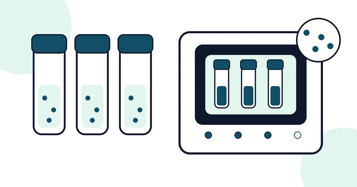
Modern blood culture systems use continuous fluorescent or colorimetric CO2 detection to monitor for microbial growth. Bottles are incubated at 35–37°C for up to 5 days (or longer if fastidious organisms are suspected). When a bottle signals positive, laboratory staff are immediately alerted. The bottle contents undergo Gram stain to give rapid morphological information (Gram-positive cocci in clusters — possible Staphylococcus; Gram-negative rods — possible Enterobacterales), and a subculture is plated.
Identification and Susceptibility Testing
Isolated organisms are identified using MALDI-TOF mass spectrometry, which provides accurate identification in minutes from a small colony. Antimicrobial susceptibility testing (AST) is performed by disk diffusion (Kirby-Bauer), automated broth microdilution (e.g. VITEK 2), or gradient strip methods (e.g. Etest). Results are reported as MIC (minimum inhibitory concentration) values and clinical breakpoints (susceptible, intermediate, resistant) according to EUCAST or CLSI guidelines. Direct susceptibility testing from positive bottles can accelerate results by 12–24 hours.
Interpreting Results: True Positive vs. Contaminant
Not all positive blood cultures represent true bacteraemia. Coagulase-negative Staphylococci (CoNS, e.g. S. epidermidis) are the commonest blood culture contaminants, originating from skin flora. A single positive bottle with CoNS in a clinically well patient is likely contamination. Two or more bottles positive with the same organism, or growth of S. aureus, Streptococcus pneumoniae, Gram-negative organisms, or Candida spp. in any bottle, is considered clinically significant. Clinical context is always required for interpretation.
References
- Public Health England. UK Standards for Microbiology Investigations: Blood Cultures. gov.uk/phe
- EUCAST. Antimicrobial susceptibility testing breakpoints. eucast.org
- Weinstein MP, et al. The clinical significance of positive blood cultures in the 1990s. Clin Infect Dis. 1997;24(4):584-602.
More questions answered
Why are blood cultures usually collected before antibiotics when possible?
Blood cultures are designed to recover viable microorganisms from the bloodstream. Starting antimicrobial treatment can reduce the number of organisms able to grow, which may lower the chance of detecting them. In a seriously ill patient, however, treatment should not be delayed unsafely just to obtain a laboratory sample. Collection timing is therefore managed by the clinical team according to the urgency of the situation.
Why are several blood culture bottles sometimes collected?
Bloodstream microorganisms may be present in low or intermittent numbers, so collecting an adequate blood volume can improve the chance of recovery. Laboratories commonly use different culture bottles to support growth under different atmospheric conditions. Multiple correctly collected samples can also help distinguish a true bloodstream infection from contamination. The exact collection procedure follows local laboratory guidance.
What causes a contaminated blood culture?
Blood culture contamination often occurs when microorganisms from the skin or collection environment enter the bottle during sampling. Contamination can create a positive result even though that organism was not genuinely circulating in the patient’s blood. Good skin preparation, aseptic technique and correct bottle handling reduce this risk. The laboratory and clinical team assess the organism, collection pattern and clinical context when deciding whether a result may represent contamination.
How long does it take for a blood culture to become positive?
An automated blood culture system continuously monitors inoculated bottles for evidence of microbial growth. Some organisms trigger a positive signal relatively quickly, while others grow more slowly or may not grow in routine culture conditions. The total incubation and reporting process depends on the laboratory method and organism, so there is no single turnaround time for every culture. Clinicians should use the local laboratory’s expected reporting schedule rather than a universal number.
What happens after a blood culture becomes positive?
When a bottle signals positive, the laboratory performs further work to identify the microorganism and assess its significance. This may include microscopy, rapid identification, subculture and antimicrobial susceptibility testing where appropriate. Important preliminary findings can be communicated before every final test is complete. The final report may therefore develop in stages rather than appearing as one result.
Can a negative blood culture rule out sepsis?
No. A negative blood culture does not rule out sepsis because infection may be located outside the bloodstream, organism numbers may be low or prior antimicrobials may reduce recovery. Some microorganisms are also difficult to grow using routine methods. Sepsis is a clinical emergency assessed using the whole clinical picture, not one culture result. Anyone suspected of having sepsis requires urgent medical assessment.
What is the difference between a blood culture and a PCR infection test?
A blood culture detects viable organisms that can grow under the laboratory’s culture conditions. PCR detects selected microbial genetic material and can be faster for particular targets, but it only finds organisms included in the assay design. Culture can provide isolates for further identification and susceptibility testing, while molecular tests answer different questions. Laboratories choose the method according to the suspected infection, specimen and clinical need.
Urine Culture vs Blood Culture: What’s the Difference and When Is Each Used?