
⚕️ Educational content only. This article explains PCR from a molecular biology and biomedical science perspective. It is not medical advice.
Polymerase chain reaction (PCR) is a fundamental molecular biology technique that amplifies specific DNA sequences from tiny starting quantities. First developed by Kary Mullis in 1983 (Nobel Prize, 1993), PCR is now indispensable in clinical diagnostics, forensic science, research, and public health. Understanding PCR underpins much of modern biomedical science.
Key Takeaways
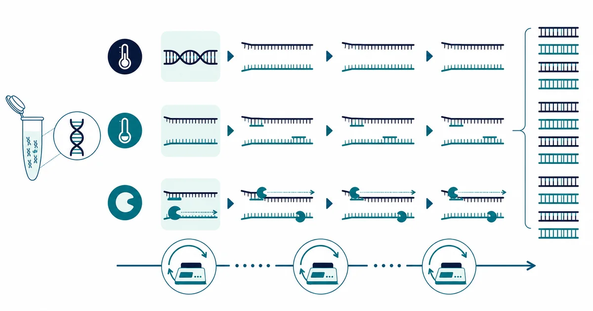
- PCR amplifies a specific DNA region using repeated cycles of denaturation, annealing, and extension.
- Thermostable Taq polymerase allows automated cycling; specific primers define the target region.
- RT-PCR converts RNA to cDNA first, enabling detection of RNA viruses (e.g. SARS-CoV-2) and gene expression analysis.
- qPCR (quantitative/real-time PCR) measures DNA quantity in real-time using fluorescent probes or intercalating dyes.
The Three Steps of PCR
Each PCR cycle consists of three steps: (1) Denaturation (94–98°C): the double-stranded DNA template is heated to separate the two strands. (2) Annealing (50–65°C): short synthetic oligonucleotide primers bind (anneal) to their complementary sequences flanking the target region on each template strand. Primer design is critical — primers are typically 18–25 bp long and selected for specific melting temperature and lack of self-complementarity. (3) Extension (72°C): Taq DNA polymerase (isolated from Thermus aquaticus, a thermophilic bacterium) synthesises new DNA from the primer, using the template strand and adding dNTPs (deoxynucleoside triphosphates). After 25–35 cycles, the target region has been amplified exponentially — over one billion copies from a single template molecule.
Key Components of a PCR Reaction
A standard PCR reaction contains: DNA template; forward and reverse primers; Taq DNA polymerase (or a higher-fidelity polymerase such as Phusion or Q5 for applications requiring accuracy); dNTPs (dATP, dCTP, dGTP, dTTP); MgCl₂ (cofactor for polymerase); and a buffer to maintain pH. The magnesium concentration is critical — too little reduces yield; too much causes non-specific amplification.
RT-PCR: Detecting RNA Targets
Reverse transcription PCR (RT-PCR) adds a step before the standard PCR: RNA is first converted to complementary DNA (cDNA) using reverse transcriptase enzyme. This allows amplification of RNA targets — crucial for detecting RNA viruses (e.g. SARS-CoV-2, influenza, HIV), for gene expression analysis (measuring mRNA levels), and for detecting alternatively spliced transcripts. The term RT-PCR is sometimes confused with real-time PCR (see below); one-step RT-PCR kits combine both the reverse transcription and PCR amplification in a single tube.
qPCR: Quantitative Real-Time PCR
Quantitative PCR (qPCR) monitors amplification in real-time using fluorescent reporters — either intercalating dyes (e.g. SYBR Green, which fluoresces when bound to double-stranded DNA) or sequence-specific probes (e.g. TaqMan probes, which are cleaved during extension to release fluorescence). The cycle threshold (Ct or Cq) is the cycle number at which fluorescence crosses a threshold — a lower Ct indicates more starting template. qPCR is used clinically to quantify viral loads (HIV RNA, HBV DNA, CMV), monitor minimal residual disease in haematological malignancies, and assess gene expression.
Clinical Applications of PCR
PCR is used across clinical microbiology and molecular diagnostics: detecting respiratory pathogens (flu, RSV, SARS-CoV-2) in nasopharyngeal swabs; diagnosing sexually transmitted infections (chlamydia, gonorrhoea, trichomonas); detecting MRSA and other resistant organisms; diagnosing genetic conditions via mutation detection; blood donor screening for HIV, HBV, HCV; and detecting BCR-ABL fusion transcript in CML for minimal residual disease monitoring. Multiplex PCR panels can simultaneously detect 15–20+ targets in a single reaction.
References
- Mullis KB. The unusual origin of the polymerase chain reaction. Sci Am. 1990;262(4):56-65.
- NIH National Human Genome Research Institute. Polymerase Chain Reaction (PCR). genome.gov
- Bustin SA, et al. The MIQE guidelines: minimum information for publication of quantitative real-time PCR experiments. Clin Chem. 2009.
More questions answered
What does PCR actually detect in a laboratory sample?
PCR detects selected nucleic-acid sequences by amplifying a defined genetic target until it can be measured. The assay only looks for the target sequence it was designed to recognise, so a negative result does not mean that every possible organism or genetic change is absent. The specimen also has to contain enough suitable nucleic acid for the method to work. Interpretation therefore depends on the assay design, specimen type and reason for testing.
What is the difference between PCR and RT-PCR?
Standard PCR amplifies DNA, while reverse-transcription PCR first converts RNA into complementary DNA before amplification. This extra step allows RNA targets, including many viral genomes or RNA transcripts, to be measured using a PCR-based workflow. The abbreviation RT-PCR is sometimes used inconsistently in general media, so the laboratory method should be checked carefully. Both methods rely on specific primers, controls and validated amplification conditions.
Why can a PCR test be negative even when the target is actually present?
A PCR result can be negative if the specimen contains too little target material, was collected at the wrong site or time, or was degraded during handling. Inhibitory substances can also interfere with amplification, depending on the specimen and assay. Laboratories use internal controls to help detect some technical problems, but no test has perfect sensitivity. A negative result therefore has to be interpreted in the context of the test and clinical question.
Can contamination cause a false-positive PCR result?
Yes. Because PCR amplification can detect very small amounts of target nucleic acid, contamination can create misleading positive signals if laboratory controls fail. Molecular laboratories separate workflow stages, use clean techniques and include negative controls to reduce this risk. Modern closed systems can further limit opportunities for amplified material to contaminate new samples. Unexpected findings may still require laboratory review or repeat testing.
What do controls do in a PCR test?
PCR runs include positive, negative and internal controls to show whether important parts of the assay are working as expected. A positive control checks that the target can be detected, while a negative control helps reveal contamination. Internal controls can help show whether extraction or amplification has been inhibited in the patient specimen. Results are not normally released if required control criteria fail.
Is a PCR cycle threshold value the same thing as the amount of target in the sample?
Not necessarily. A cycle threshold value reflects when an amplification signal crosses a defined detection threshold within a particular PCR system. It can relate to the amount of target present, but values are affected by assay design, specimen quality, extraction method and instrument settings. Ct values are therefore not automatically comparable between different assays or laboratories. They should only be interpreted according to the validated method and reporting guidance used by that laboratory.
How is PCR different from an antigen test?
PCR detects selected genetic material, while an antigen test detects particular proteins from the target organism or substance. PCR often includes an amplification step that can make it analytically more sensitive, although performance depends on the specific assay and specimen. Antigen tests can be faster and simpler in some settings but answer a different analytical question. The appropriate method depends on the purpose of testing rather than one technology always being better.
PCR vs Antibody Test: What’s the Difference and Which One Do You Need?